Phytogenics and its effect on E. coli adhesion

Plant derived products and extracts can have a positive impact on animal’s health and the immune system. Stefanie Gärtner and collegues looked at the effects of phytogenic additives on the adhesion of pathogenic E.coli to porcine intestinal cells and on immunological parameters in piglets.
By Stefanie Gartner
Although there is evidence that phytogenics can have an effect on the immune system it still seems difficult to assess the efficacy of plants or plant extracts due to a lack of targeted studies and due to the situation, that many studies have used mixtures of different plant derived products. Generally, the mode of action and the functional aspects of phytogenic feed additives still need further studies in farm animals.
The hypothesis of the study presented here is, that a phytogenic product (Fresta® F, hereafter called FF) can interact with the intestinal microbiota and with the immune reaction of piglets and stabilize intestinal and general health. The aim of the study presented here was to investigate the impact of phytogenic compounds under in vitro conditions regarding the potential impact on a pathogenic Escherichia coli strain. The interference of the plant derived product with a) the adhesion of pathogenic E. coli to IPEC-J2 cells, a permanent porcine jejunal cell line, b) the binding of pathogenic E. coli to the plant product itself and c) the potential inhibitory activity on bacterial growth were studied under in vitro conditions. A feeding trial was performed to assess the potential impact of the phytogenic feed additive on immune reactions in piglets. Therefore the impact of the plant derived products on the phagocytosis activity and several surface markers of leukocytes in the peripheral blood was investigated.
In vitro testing
Pathogenic E. coli adhesion on porcine intestinal cells was tested using IPEC-J2 cell-line. The amount of 1×10⁵ bacteria/ml medium was incubated with these cells for 90 minutes without or FF (1%). Binding of the E. coli strain to the epithelial cells was visualized after detachment of the epithelial monolayer from the culture vessels. The cell suspension was characterized in a flow cytometer assessing the number of fluorescent epithelial cells, considered as cells with attachment of E. coli, and the fluorescence intensity (FACS Calibur, BD, USA). Adhesion of pathogenic E. coli to the phytogenic product: Microtiter-plates were coated with a 1%-solution of FF or with bovine serum albumin. The wells were washed to remove unbound material and incubated with the pathogenic E. coli strain (1×10⁷/ml) for 30 minutes at 22°C to allow bacterial adhesion to the coated or non-coated wells.
The removal of non-adhering bacteria was done in a washing step.
After addition of nutrient broth, the wells were incubated for 24 h. The bacterial growth was considered to be influenced by the number of attached bacteria indicating the potential of the plant product to bind the tested pathogenic strain. The estimation of bacterial growth was done semi quantitatively at 620 nm by photometry/turbidometry. The pathogenic E. coli-strain (1×10⁷/ml) was cultured with different concentrations (1%, 0.5%, 0.25%, 0.125%, 0.06%, 0.03%) of the phytogenic product at 36°C- 38°C over 24 h. the bacterial growth curves were measured semi quantitatively by photometry/turbidometry at 620nm.
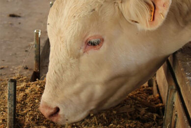
Feeding trial with piglets
A feeding trial was performed with four groups of 12 castrated male piglets each. Weaning age was 21 days and the animals were kept for four weeks. The diet was the complete starter diet covering the nutrient requirements (Table 1). The control group received the diet without addition of FF, two groups received the diet with FF at 400 ppm and 2000 ppm and the fourth group received the diet with a galactomannan powder (28 %) at an inclusion level of 1500 ppm. The animals were sacrificed after 28 days for the assessment of the immune response. The immunological traits in the blood were phagocytosis activity of peripheral granulocytes and monocytes (PHAGO-Test®, ORPEGEN Pharma GmbH, Germany), the assessment of the leukocyte surface markers (flow cytometry, FACS Calibur, Fa. BD, USA) by a panel of porcine specific antibodies: SIRPα (SWC3a, myeloid cells), CD2 (T cells), CD4 (helper T cells), CD8β (cytotoxic T cells and memory helper cells), CD8α (cytotoxic T cells, NK cells and subpopulation of γδ T cells), CD5 (T cells and B cell subpopulation), TCR1 (γδT cells), CD25 (IL-2 receptor, marker for T cell activation; CD25high marker for regulatory T cells), CD45RC (reduced expression indicates cell activation) CD21 (B-cells) , CD14 (mononuclear phagocytes) MHCII (B cells, dendritic cells, porcine TCR alpha/beta CD8+ T cells, mononuclear phagocytes). The proliferation-rate of peripheral lymphocytes tested with different mitogens (Concanavalin A, Pokeweed mitogen, Phytohaemagglutinin). In the proximal jejunum, intraepithelial leukocytes were isolated and leukocyte surface markers were measured by flow cytometry.
Results and discussion
The in vitro data indicated a decreased adhesion of the pathogenic E. coli strain to intestinal epithelial cells when 1% of the phytogenic product was added (Figure 1). The intensity of fluorescence increased by a factor of two in the control experiments compared to the FF group, which showed almost constant fluorescence intensity. This could be interesting because lowered adhesion capacities of pathogenic bacteria to the intestinal epithelium might decrease the risk for gastrointestinal disorders in piglets. The other in vitro data indicated that the phytogenic product had some binding capacity for the pathogenic E. coli-strain while there was no direct inhibitory effect on the growth of the bacteria under the given experimental conditions. Therefore, the potential interference of the phytogenic product with the bacteria-host interaction seems to be the most relevant outcome of the in vitro studies and indicates a potential impact of the product on the intestinal bacterial host-interaction. The in vivo studies followed the concept of a conventional feeding trial. The animals did not show any signs of diarrhoea or digestive disorders during the trial period. The phagocytosis activity of peripheral granulocytes and monocytes was not affected by the different treatments after 28 days. The different cell surface markers that were used as indicators of potential treatment effects on the leukocyte differentiation and function were relatively constant, except for the CD8 negative γδ T cells and MHCII+ CD5- cells (Table 2; Figures 2 and 3). MHC class II is constitutively expressed on B cells and dendritic cells. Furthermore its expression on mononuclear cells is positively regulated by IFN-γδ and negatively regulated by IL-10. (In addition porcine TCR alpha/beta CD8+ T-cells do express MHC class II. However, those cells are positive for CD5.) The differences in the percentage of γδ-T-cells might be interesting due to their importance in young animals, acting probably as regulatory cells in first-line defense situation. The physiological function of those cells is not completely clear yet and it can be expected, that their role in the immune reaction has been underestimated in the past. The number of MHCII+ CD5- cells in the peripheral blood was decreased in the group fed the galactomannan powder compared to the control group. This may indicates that the dietary treatments had an impact on the antigen presenting cells (macrophages and/or dendritic cells), which are relevant for the initiation of immune responses. The proliferation activities of peripheral blood lymphocytes after stimulation with the three mitogens (Con A, PHA, PWM) were not different between the test groups. There were also no differences in the expression of surface markers of the intraepithelial leukocytes in the jejunum of the piglets.
Conclusion
The results from the in vitro trials indicate an interference of the phytogenic feed additive with the adhesion of E. coli to IPEC-J2 cells. If this effect could be linked to the binding capacity of the product itself or to its inhibitory effects on the tested pathogenic E. coli-strain has to be further investigated. The feeding trial showed some differences in the immunological traits between the groups. The enhancement of the γδ T cells and the subsidence of the MHCII+CD5- cells, probably macrophages or dendritic cells, are interesting due to their involvement in the initiation and regulation of the immune response. Further studies should be done to explain the consequence for animal health.
Source: FeedMix vol 18 nr 1, 2010
Join 26,000+ subscribers
Subscribe to our newsletter to stay updated about all the need-to-know content in the feed sector, three times a week.